41 eye diagram with labels and functions
Parts of Human Eye and Their Functions - MD-Health.com Eye Parts. Description and Functions. Cornea. The cornea is the outer covering of the eye. This dome-shaped layer protects your eye from elements that could cause damage to the inner parts of the eye. There are several layers of the cornea, creating a tough layer that provides additional protection. These layers regenerate very quickly, helping ... Eye Anatomy: 16 Parts of the Eye & Their Functions The following are parts of the human eyes and their functions: 1. Conjunctiva The conjunctiva is the membrane covering the sclera (white portion of your eye). The conjunctiva also covers the interior of your eyelids. Conjunctivitis, often known as pink eye, occurs when this thin membrane becomes inflamed or swollen.
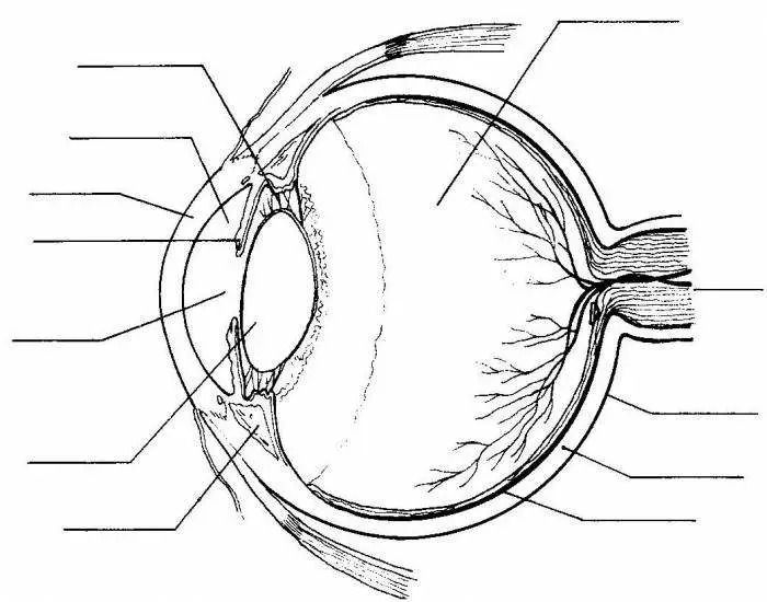
Label the Eye - The Biology Corner Label the Eye Shannan Muskopf December 30, 2019 This worksheet shows an image of the eye with structures numbered. Students practice labeling the eye or teachers can print this to use as an assessment. There are two versions on the google doc and pdf file, one where the word bank is included and another with no word bank for differentiation.

Eye diagram with labels and functions
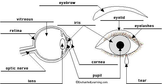
Anatomy of the Eye Diagrams for Coloring/Labeling, with ... This printable contains 13 clear and simple cross sectional diagrams of the human eye. They photocopy well and are great for use as a labeling and coloring exercise for your students. The core eye anatomy diagram, designed as the labeling exercise, has a fully colored and labeled reference chart to go with it. Eye Anatomy Diagram - EnchantedLearning.com Retina - light-sensitive tissue that lines the back of the eye. It contains millions of photoreceptors (rods and cones) that convert light rays into electrical impulses that are relayed to the brain via the optic nerve. Rods - cells the in the retina that sense brightness (they are photoreceptors). Night vision involves mostly rods (not cones). Labeled Eye Diagram - Science Trends What you want to interpret as a major part of the human eye is somewhat up to the individual, but in general there are seven parts of the human eye: the cornea, the pupil, the iris, the lens, the vitreous humor, the retina, and the sclera. Let's take a closer look at each of these components individually. The Cornea
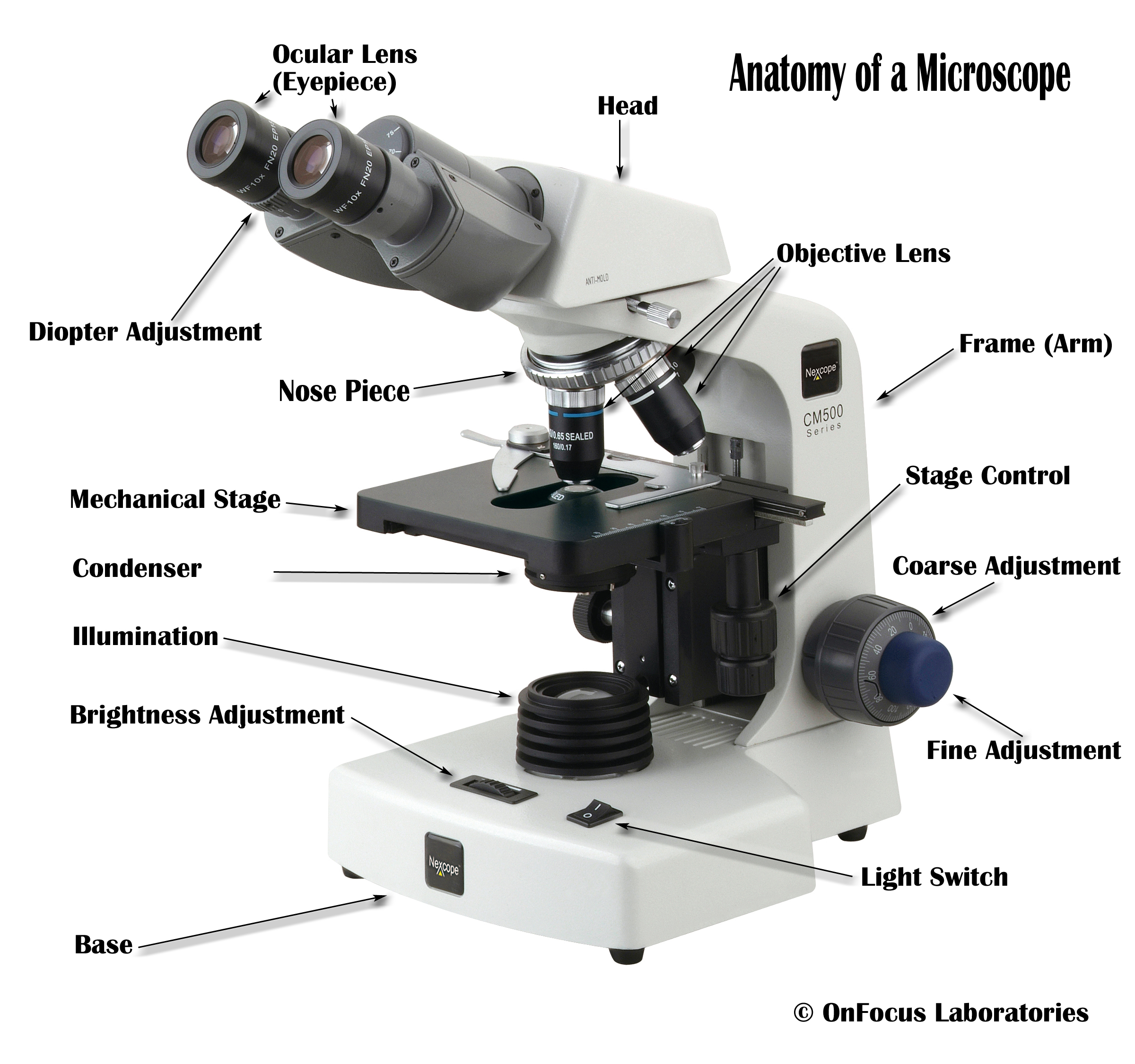
Eye diagram with labels and functions. Microscope Parts and Functions With Labeled Diagram and ... Microscope Parts and Functions With Labeled Diagram and Functions How does a Compound Microscope Work?. Before exploring microscope parts and functions, you should probably understand that the compound light microscope is more complicated than just a microscope with more than one lens.. First, the purpose of a microscope is to magnify a small object or to magnify the fine details of a larger ... Generate eye diagram - MATLAB eyediagram eyediagram (x,n,period) sets the labels on the horizontal axis to the range between - period /2 to period /2. eyediagram (x,n,period,offset) specifies the offset for the eye diagram. The function assumes that the ( offset + 1)th value of the signal and every n th value thereafter, occur at times that are integer multiples of period. Microscope, Microscope Parts, Labeled Diagram, and Functions Microscope, Microscope Parts, Labeled Diagram, and Functions What is Microscope? A microscope is a laboratory instrument used to examine objects that are too small to be seen by the naked eye. It is derived from Ancient Greek words and composed of mikrós, "small" and skopeîn,"to look" or "see". The Eyes (Human Anatomy): Diagram, Function, Definition ... Eye Conditions. Age-related macular degeneration: Causes loss of central vision as you get older.. Amblyopia: Often called lazy eye, this condition starts in childhood.One eye sees better than the ...
Human Eye Diagram, How The Eye Work -15 Amazing Facts of Eye First, light rays enter the eye through the cornea, the clear front "window" of the eye. The dome shaped cornea bends light to help the eye focus. From the cornea, the light passes through an opening called the pupil. The amount of light passing through is controlled by the iris, or the colored part of your eye. Eye Parts Labeling and Functions Flashcards - Quizlet layer of cells on the back of the eye cornea function helps protect the eye, and bends light to make an image appear on the retina through the lens iris function controls how much light enters the eye lens function makes an image on the eye's retina and can focus on objects that are close and far away by changing shape optic nerve function Eye Diagram Teaching Resources | Teachers Pay Teachers The Human Eye Overview Reading Comprehension and Diagram Worksheet by Teaching to the Middle 58 $1.50 Zip This passage briefly describes the human eye (900-1000 Lexile). 14 questions (matching and multiple choice) assess students' understanding. Students label a diagram of 6 parts of the eye. I've included a color and BW version, as well as a key. Structure and Functions of Human Eye with labelled Diagram Structure and Functions of Human Eye with labelled Diagram Biology Biology Article Structure Of Eye Structure of the Eye The eye is one of the sensory organs of the body. In this article, we shall explore the anatomy of the eye The structure of the eye is an important topic to understand as it one of the important sensory organs in the human body.
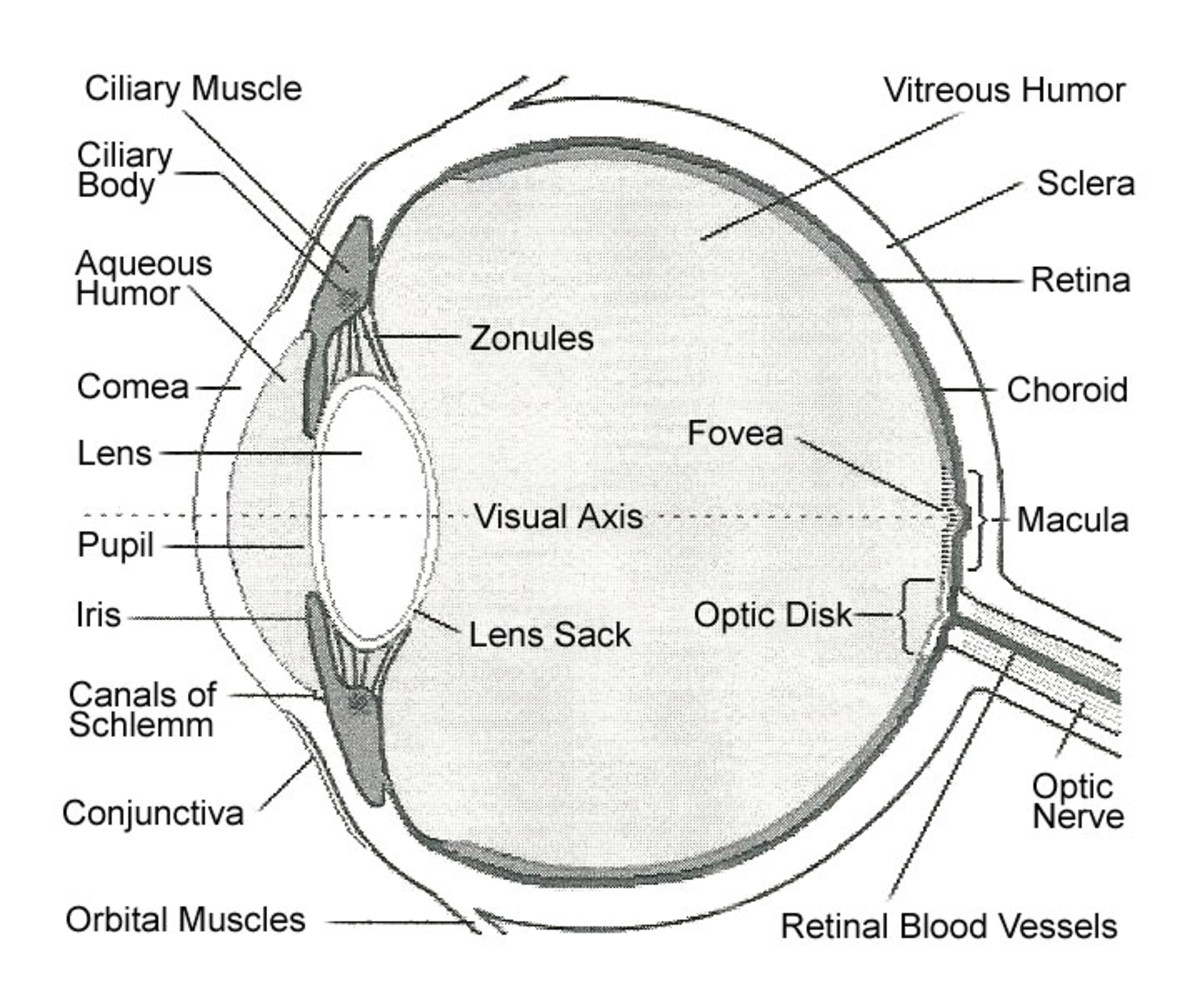
Human Eye: Structure of Human Eye (With Diagram) | Biology The human eye is a very sensitive and delicate organ suspended in the eye socket which protects it from injuries. It essentially consists of CORNEA, LENS & RETINA besides many other parts such as Iris, Pupil and aqueous humour, vituous humour etc. Each one has got a specific function. A section of the eye is as shown in Fig. 2.2. ADVERTISEMENTS: Eye Diagram With Labels and detailed description A brief description of the eye along with a well-labelled diagram is given below for reference. Well-Labelled Diagram of Eye The anterior chamber of the eye is the space between the cornea and the iris and is filled with a lubricating fluid, aqueous humour. The vascular layer of the eye, known as the choroid contains the connective tissue. Structure of Human Eye (With Diagram) | Human Body ADVERTISEMENTS: In this article we will discuss about the structure of human eye. Structure of Human Eye: The eye is a hollow, spherical structure measuring about 2.5 cm in diameter. ADVERTISEMENTS: Its wall is composed of three coats: 1. The outer fibrous coat— sclera, cornea. 2. The middle vascular coat— choroid, ciliary body, iris. 3. […] Draw a labeled diagram of human eye. Write the functions ... Cornea of the eye is the first sight where convergence of light rays takes place. Iris is that part of the eye which controls the amount of light entering the eye through the pupil. Pupil is a type of small hole through which light enters the eye. The eye lens is a convex lens just behind the pupilwhich converges the light rays towards the ratina.
Labeled Eye Diagram - Science Trends What you want to interpret as a major part of the human eye is somewhat up to the individual, but in general there are seven parts of the human eye: the cornea, the pupil, the iris, the lens, the vitreous humor, the retina, and the sclera. Let's take a closer look at each of these components individually. The Cornea
Eye Anatomy Diagram - EnchantedLearning.com Retina - light-sensitive tissue that lines the back of the eye. It contains millions of photoreceptors (rods and cones) that convert light rays into electrical impulses that are relayed to the brain via the optic nerve. Rods - cells the in the retina that sense brightness (they are photoreceptors). Night vision involves mostly rods (not cones).
Anatomy of the Eye Diagrams for Coloring/Labeling, with ... This printable contains 13 clear and simple cross sectional diagrams of the human eye. They photocopy well and are great for use as a labeling and coloring exercise for your students. The core eye anatomy diagram, designed as the labeling exercise, has a fully colored and labeled reference chart to go with it.


Post a Comment for "41 eye diagram with labels and functions"