41 ribosome diagram with labels
Ribosome Images - University of California, Santa Cruz Ribosome Images. All fullsize images are 300 pixels/inch and suitable for high resolution reproduction. All TIFF images use the CMYK color gamut, are are LZW compressed. All JPEG images use the RGB color gamut and have minimal compression. Solved The ribosome in the diagram is in the process of - Chegg The ribosome in the diagram is in the process of synthesizing a protein using directions transcribed from the DNA. Use the labels to identify each of the structures involved in translation and protein synthesis. Question: The ribosome in the diagram is in the process of synthesizing a protein using directions transcribed from the DNA.
Ribosomes- Definition, Structure, Functions and Diagram Ribosomes Definition The ribosome word is derived - 'ribo' from ribonucleic acid and 'somes' from the Greek word 'soma' which means 'body'. Ribosomes are tiny spheroidal dense particles (of 150 to 200 A0 diameters) that are primarily found in most prokaryotic and eukaryotic. They are sites of protein synthesis.
Ribosome diagram with labels
Ribosomes | Definition, Examples, Diagrams - Toppr Click here to learn the concepts of Ribosomes from Biology. ... Draw a diagram of an animal cell and label at least eight organelles in it. A Labelled Diagram Of Mitochondria with Detailed Explanation - BYJUS It is a viscous or a gel-like fluid containing a mixture of enzymes, ribosomes, inorganic ions, mitochondrial DNA, nucleotide cofactors, and organic molecules. It is involved in the cellular respiration and production of ATP molecules. Cristae The inner layer, surrounded by the folds of the mitochondrial matrix are collectively referred to Cristae. Two-Photon Excitation Microscopy for the Study of Living ... With increasing depth, it becomes increasingly difficult to introduce fluorescent labels into the cells and tissue of interest. The use of tissue-specific fluorescent protein expression in transgenic animals enhances the usefulness of two-photon excitation in vivo imaging, allowing efficient labeling of tissues at any depth.
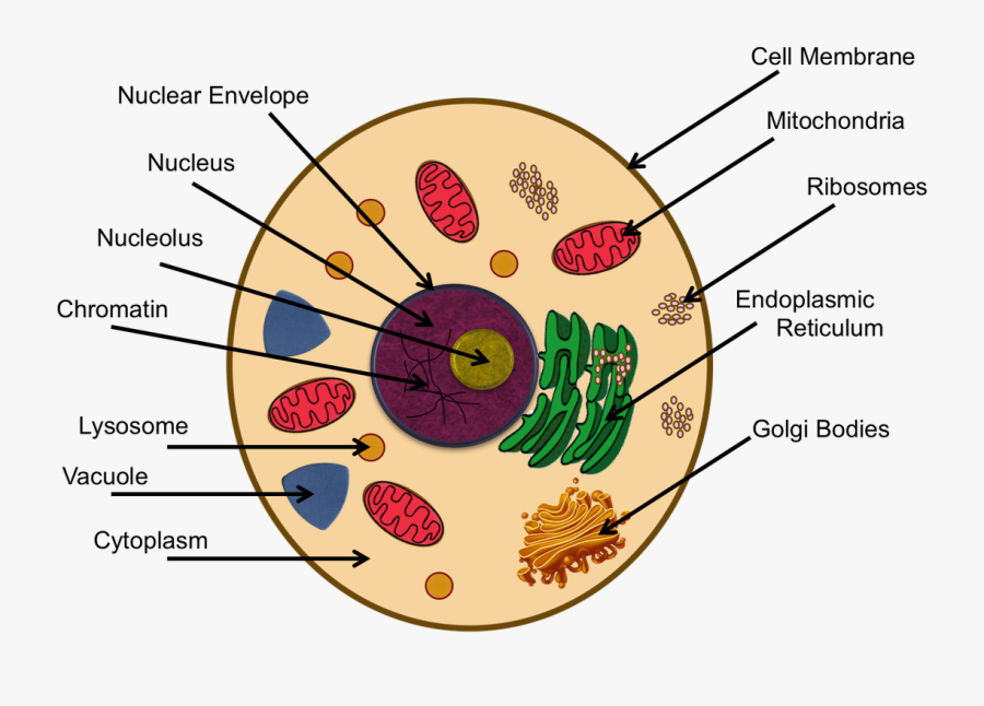
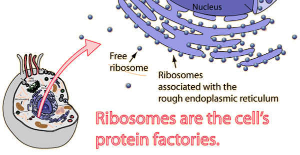
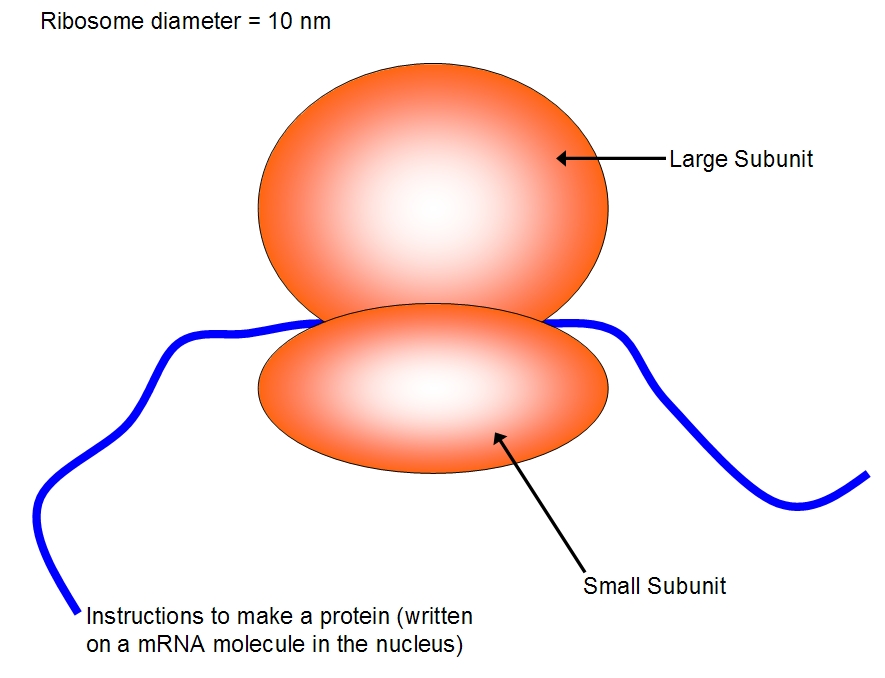
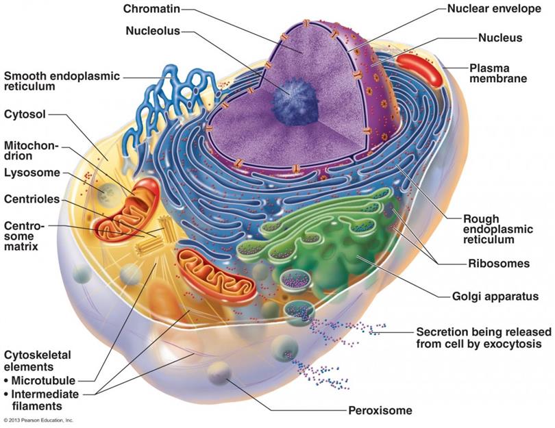
Ribosome diagram with labels. A Labeled Diagram of the Animal Cell and its Organelles A Labeled Diagram of the Animal Cell and its Organelles. There are two types of cells - Prokaryotic and Eucaryotic. Eukaryotic cells are larger, more complex, and have evolved more recently than prokaryotes. ... Ribosomes are small, spherical organelles comprising 65% ribosomal RNA and 35% ribosomal proteins. Animal cells contain ribosomes with ... Animal Cell Diagram with Label and Explanation: Cell ... - Collegedunia Animal cell is a typical Eukaryotic cell enclosed by a plasma membrane containing nucleus and organelles which lack cell walls, unlike all other Eukaryotic cells. The typical cell ranges in size between 1-100 micrometers. The lack of cell walls enabled the animal cells to develop a greater diversity of cell types. What Are Ribosomes? - Definition, Structure and its Functions - BYJUS A ribosome is a complex molecular machine found inside the living cells that produce proteins from amino acids during a process called protein synthesis or translation. The process of protein synthesis is a primary function, which is performed by all living cells. Ribosomes are specialized cell organelles and are found in both prokaryotic and ... Labeled Plant Cell With Diagrams | Science Trends The ribosomes are created in the nucleolus of the cell. Ribosomes are made out of two smaller subunits, a large ribosomes subunit and a small ribosomal subunits. The transfer RNA or tRNA encodes the correct series of genetic instructions into the mRNA or messenger RNA, which is what ensures that the right proteins are created.
Mastering Quiz: Chapter 7A Microbial Genetics Flashcards | Quizlet c. mRNA binds to a ribosome in the cytoplasm. d. A molecule of RNA is formed based on the sequence of nucleotides in DNA. ... Drag the correct labels under the diagrams to identify the events of RNA processing. Drag the labels onto the diagram to identify how nucleotides pair up. Labels can be used once, more than once, or not at all. Nucleotide Structure: DNA Diagram | Science Trends Ribosomal RNA, as the name implies, is involved in the creation of ribosomes, and it comprises around 60% of the mass of ribosomes. This form of RNA is needed to properly align the mRNA and give the mRNA a point of attachment. Transfer RNA brings the requisite amino acids to the ribosomes so that they can be used to synthesized proteins. Bacteria in Microbiology - shapes, structure and diagram - Jotscroll Bacterial spores. Bacterial endospores layers. Bacteria cells are the smallest living cells that are known; even though viruses are smaller than bacteria, viruses are not living cells. There are different types of bacteria with various sizes, shapes, and structures. The bacteria shapes, structure, and labeled diagrams are discussed below. Solved In the following diagram of a ribosome, assign the - Chegg In the following diagram of a ribosome, assign the correct labels. Who are the experts? Experts are tested by Chegg as specialists in their subject area. We review their content and use your feedback to keep the quality high. Transcribed image text: In the following diagram of a ribosome, assign the correct labels.
Ribosomes Images Stock Photos, Pictures & Royalty-Free Images - iStock Ribosomes vector illustration. Anatomical and medical labeled scheme with tRNA, Amino acid, protein, cell, small and large subunit, mRNA. Explained closeup diagram. ribosomes images stock illustrations DNA Labeling: Transciption and Translation - The Biology Corner This worksheet shows a diagram of transcription and translation and asks students to label it; also includes questions about the processes. Name: _____ ... How does the ribosome know the sequence of amino acids to build? 12. What is the difference between a codon and an anticodon? PDF Quick Review Transcription and Translation - WPMU DEV label the diagram. 2. ... 910dnamrnait carries the genetic code from dna to ribosome to make a proteinit carries the amino acids to make proteinbecause the genetic code is the recipe to make a protein and is contained in a mrnacodons are in mrna and anti codons are groups of 3 bases in trnatranscription takes place in nucleus; translation takes ... Animal Cell Diagram Ribosomes Simple : Functions and Diagram Each ribosome is made up of two subunits i. e large subunit and small subunit with their own distinct shapes. They are situated in the cytosol, some bound and free-floating to the membrane of the coarse endoplasmic reticulum. Ribosomes were first described by George E.
Clip Art Ribosomes Diagram - Cytoskeleton In Animal Cell Project , Free Transparent Clipart ...
Prokaryotic Cells - BioNinja Ribosomes – complexes of RNA and protein that are responsible for polypeptide synthesis (prokaryote ribosome = 70S) Cell membrane – Semi-permeable and selective barrier surrounding the cell Cell wall – rigid outer covering made of peptidoglycan; maintains shape and prevents bursting (lysis)
Anatomy & Physiology I Chapter 4 Flashcards | Easy Notecards
Ribosome - Wikipedia The ribosomal proteins and rRNAs are arranged into two distinct ribosomal pieces of different sizes, known generally as the large and small subunit of the ribosome. Ribosomes consist of two subunits that fit together (Figure 2) and work as one to translate the mRNA into a polypeptide chain during protein synthesis (Figure 1).

Science(Claudia Aguilera): Vocabulary #2
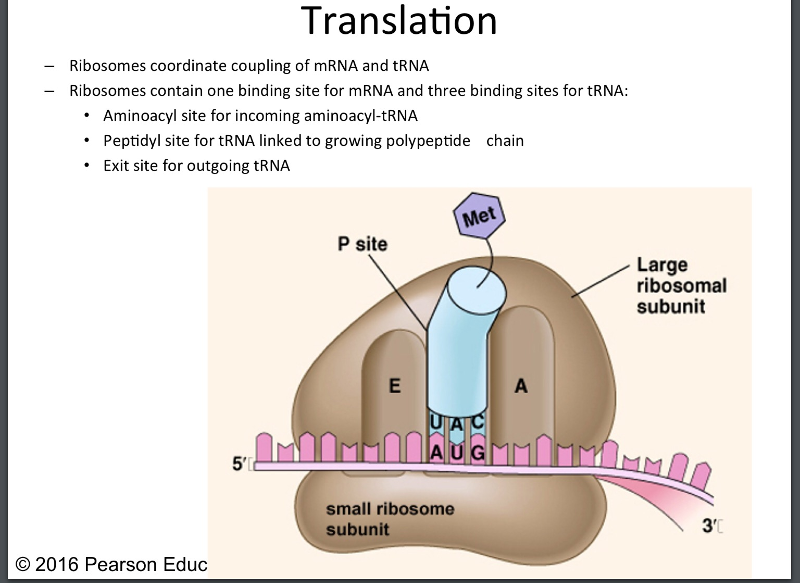
Bio 1113 - Unit 11 - Gene Expression Flashcards | Quizlet In the following diagram of a ribosome, assign the correct labels: Label 1: a tRNA attached to a polypeptide is found in this area of the ribosome Label 2: a tRNA attached to a single amino acid enters here Label 3: a tRNA that is not attached to anything exits here Label 4: a tRNA molecule Label 5: growing polypeptide Label 6: mRNA being ...

Because So Much is Riding on your Ribosomes!
Cell Organelles- Definition, Structure, Functions, Diagram In the case of prokaryotic cells, the ribosomes are of the 70S with the larger subunit of 50S and the smaller one of 30S. Eukaryotic cells have 80S ribosomes with 60S larger subunit and 40S smaller subunit. Ribosomes are short-lived as after the protein synthesis, the subunits split up and can be either reused or remain broken up.
Ribosome Explained By Analogy Metaphor Examples
Schematic diagram of ribosome biogenesis. In the ... A comparative quantitative label-free proteomic analysis revealed that a total of 215 proteins were differentially accumulated between the young siliques of the ...
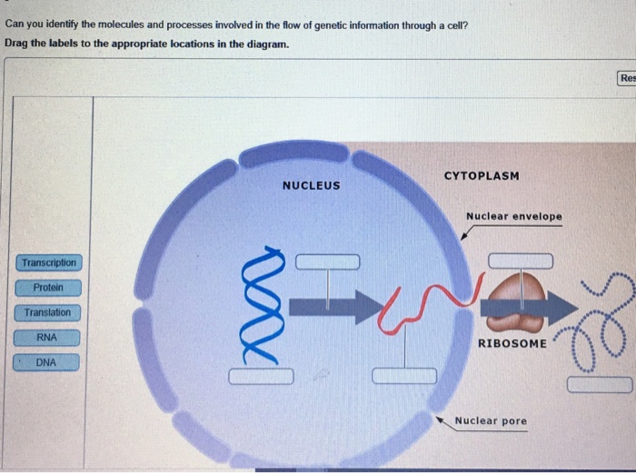
Solved: Can You Identify The Molecules And Processes Invol... | Chegg.com
Animal Cells: Labelled Diagram, Definitions, and Structure - Research Tweet Ribosomes Ribosomes create proteins. They can float freely in the cytoplasm or can be attached to the nuclear envelope. They create proteins by assembling amino acids into polypeptides. As the ribosomes build an amino acid chain, the chain is pushed into the endoplasmic reticulum.
Topic 1.2 Ultra-Structure of Cells - AMAZING WORLD OF SCIENCE WITH MR. GREEN
protein synthesis diagram labeled - TheFitnessManual Switch RNAs (tRNAs) deliver amino acids to the ribosome. - "protein synthesis diagram labeled" tRNAs are additionally RNA polymers. They're typically between 75 and 90 RNA nucleotides lengthy. However in contrast to mRNAs, that are linear, hydrogen bonding between nucleotides inside a tRNA causes it to fold up.
Animal Cell Features
Ribosomes - Definition, Structure, Size, Location and ... Ribosomes are made of proteins and ribonucleic acid (abbreviated as RNA), in almost equal amounts. It comprises of two sections, known as subunits. The tinier ...

The structure of the ribosome Infographics. illustration. #Sponsored , #ad, #ribosome#structure# ...
Active Ribosome Profiling with RiboLace - PubMed Ribosome profiling, or Ribo-seq, is based on large-scale sequencing of RNA fragments protected from nuclease digestion by ribosomes. Thanks to its unique ability to provide positional information about ribosomes flowing along transcripts, this method can be used to shed light on mechanistic aspects … Active Ribosome Profiling with RiboLace

Structure of the Mammalian 80S Ribosome at 8.7 Å Resolution: Structure
Lineage tracing reveals the phylodynamics, plasticity, and ... May 26, 2022 · (J) Venn diagram illustrating the classification of expansions to gene modules based on a p value threshold of 0.05 using a permutation test against nonexpanding background. Combining the aforementioned single-cell fitness scores with single-cell transcriptomes for each tumor, we next identified genes associated with changes in fitness for each ...

Microbe Notes | Online Microbiology and Biology Notes
Ribosome - Definition, Function and Structure | Biology Dictionary A. Ribosomes translate the 4 base language of DNA into the 20 base language of proteins, allowing for many more combinations. B. The 4 different nucleobases of DNA can be recombined endlessly to produce new proteins. C. Ribosomes can modify proteins with carbohydrates to make them unique. Answer to Question #2 3.
Print Usc bridge nurs 500 3.5 Translation flashcards | Easy Notecards
Structure of Ribosome (With Diagram) - Biology Discussion A bacterial ribosome is about 250 nm in diameter and consists of two subunits, one large and one small. Both subunits consist of one or more molecules of rRNA and an array of ribosomal proteins. ADVERTISEMENTS: Association of two subunits is called mono-some. The structure of prokaryotic ribosome is given in the figure 8.2 B.

In The Following Diagram Of A Ribosome Assign The Correct Labels - Atkinsjewelry
Nucleus and ribosomes (article) | Khan Academy Structure and function of the nucleus and ribosomes of a cell. ... (In the first diagram in this article, the red dots represent bound ribosomes; ...

Biology matters: Clarification: Events at the rER (synthesized protein will end up in the rER ...
Genetics: A Conceptual Approach - Solutions and Problem ... Enter the email address you signed up with and we'll email you a reset link.

AP Biology Unit 2: The Cell Flashcards | CourseNotes
Ribosomes: Structure, Composition, and Assembly (With Diagram) Ribosomes in the cytoplasm of eukaryotic cells have a sedimentation coefficient of about 80 S (MW about 4.5 x 10 6) and are composed of 40 S and 60 S subunits. In prokaryotic cells, ribosomes are typically about 70 S (MW about 2.7 x 10 6) and are formed from 30 S and 50 S subunits.

ribosome
Ribosome - protein factory - definition, function, structure and biology The protein translation by a ribosome consists of three stages: (1) Initiation, (2) Elongation, and (3) Termination. Initiation - the ribosome assembles around the target mRNA. A small ribosome subunit links onto the "start-end" of an mRNA strand. "Initiator tRNA" also enters the small subunit and binds to the start codon (most commonly, AUG).
Plant and Animal Cell Diagram - Plant and Animal cells
Ribosome and protein synthesis, diagram - Stock Image - C029/3020 Diagram showing protein synthesis in cells (translation). Messenger ribonucleic acid (mRNA, blue with coloured nucleotides) is read by a ribosome (pink). The molecules of transfer RNA (tRNA, key-shaped) each bring an amino acid (orange dot) to bind to the ribosome's protein synthesis site.
Post a Comment for "41 ribosome diagram with labels"